
October 17, 2019
Salk mouse synthetic embryo model could open new avenues for understanding early development, among many other applications for human health
Salk synthetic embryo model could open new avenues for understanding early development, among many other applications for human health
LA JOLLA—Although graduating from school, a first job and marriage can be important events in life, some of the most significant events happen far earlier: in the first few days after a sperm fertilizes an egg and the cell begins to divide.
The way the first 100 cells (collectively called a blastocyst) organize themselves has profound implications for whether a pregnancy is successful, how organs form, and potentially even for diseases later in life, such as Alzheimer’s. However, scientists have not had a good way to model how a blastocyst is formed, until now.
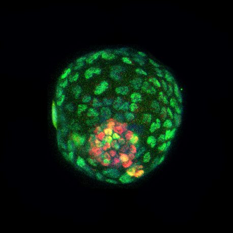
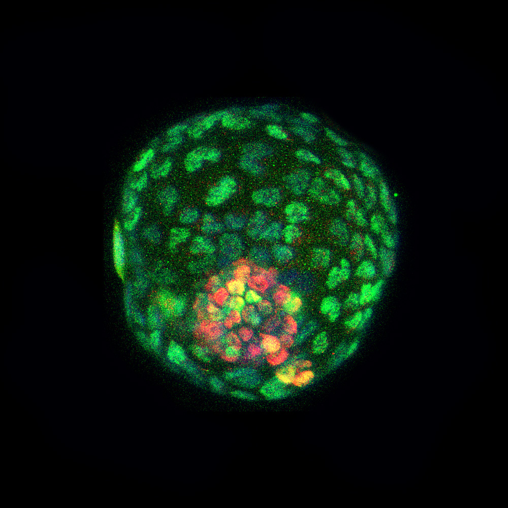
Click here for a high-resolution image.
Credit: Salk Institute/Waitt Advanced Biophotonics Core Facility
For the first time, researchers at the Salk Institute and the University of Texas Southwestern Medical Center have created mouse blastocyst-like structures, or “blastoids,” from a single cultured cell, circumventing the need for natural embryos. As they reported October 17, 2019, in the journal Cell, these cultured blastoids have the same structure as natural blastocysts and can even implant in the uterus—and could help advance research into development as well as inform issues around pregnancy, infertility, or health problems later in the offspring’s life.
“These studies will help us to better understand the very beginnings of life; how early on in life a single cell can give rise to millions of cells and how they are assembled in space and time to give rise to a fully developed organism. Importantly, this work avoids the use of natural embryos and is scalable,” says Juan Carlos Izpisua Belmonte, a professor in Salk’s Gene Expression Laboratory.
Natural blastocysts, which can become an embryo once they implant in a uterus, have proven difficult to study. The problem is that animal models, such as mice, only produce these structures in small numbers, and scientists cannot easily test the effects of malnutrition or exposure to toxins or a variety of genetic mutations on development at a level sufficient for study.
“We are optimistic that this work will enable important research into early developmental defects,” says Assistant Professor Jun Wu of UT Southwestern, who co-led the study.
The Salk and UT Southwestern teams developed the blastoids using embryonic and, more importantly, adult mouse cells. The adult cells were put into a chemical solution that prompted them to turn into induced pluripotent stem cells, or iPSCs, which can turn into almost any kind of tissue in the body.
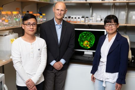
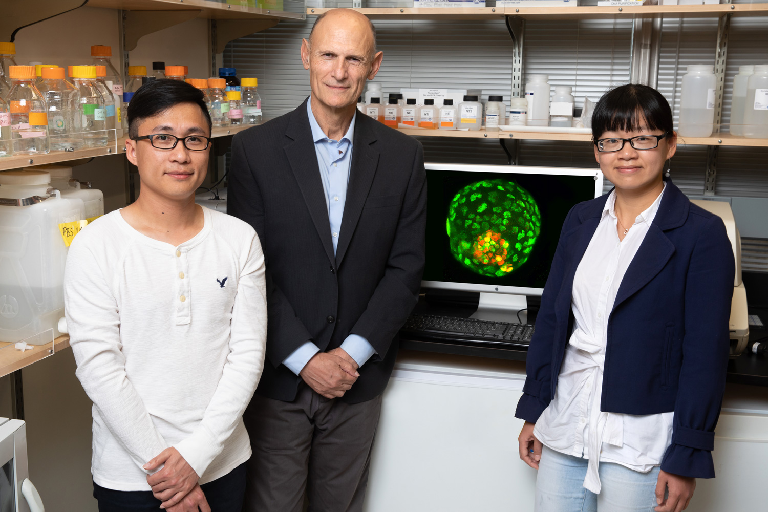
Click here for a high-resolution image.
Credit: Salk Institute
To encourage the iPS cells to form blastoids, the researchers put them in small groups in a special culture medium where they soon formed connections with each other. This was exactly what the researchers hoped to see—the cells were beginning to form structures similar to the developmental stage before a fertilized egg becomes a blastocyst.
Over time, the connected cells started forming a ball with an inner and outer layer. The cells facing inward accumulated proteins that made them distinct from the outside cells. The cells facing outward also began activating a protein called YAP, which entered the cell nucleus and began the process of inducing expression of proteins to build what could eventually become a placenta.
“The formation of blastoids mimics the natural developmental process,” says Ronghui Li, co-first author of the study and a postdoctoral fellow in the Izpisua Belmonte lab.
The blastoids contained the same three primordial cell types (from which all the cells of an adult organism come) found in natural blastocysts. They were also a similar size as natural blastocysts and showed a similar gene signature. Further experiments indicated that the blastoids could further develop into structures resembling early post-implantation embryos.
“I think this kind of resource is going to be very powerful for studying early development in mammals,” says Cuiqing Zhong, co-first author of the study and a postdoctoral fellow in the Izpisua Belmonte lab.
The team next plans to use gene-editing tools to understand how genetic changes in blastoids affect the three different cell types. The blastoids also provide a new model for testing drugs and chemicals for future therapies.
The blastoids cannot yet develop into functional embryos; instead, the cells grow into disorganized tissue. But the scientists believe the blastoids may reveal details on later stages of embryonic development.
“With further optimization, this technology might lead to the generation of fully functional blastoids able to develop up to the stages when different organ primordia are formed and thus be the seeds for organoids that could be used as invaluable sources for organ transplantation,” adds Belmonte.
“The noted physicist Richard Feynman once said, ‘What I cannot create, I do not understand.’ How life starts from a fertilized egg still remains a mystery, and our blastoid approach will help researchers gain novel insights into this process.” says Wu.
Other authors included Yang Yu (co-first author) of Salk and Peking University Third Hospital; Haisong Liu, Lei Shi, Yuta Takahashi, Hsin-Kai Liao and Concepcion Rodriguez Esteban of Salk; Zheying Min and Jie Qiao of Peking University Third Hospital; Masahiro Sakurai and Leqian Yu of UT Southwestern; Yulei Wei of UT Southwestern and Wuyi University; and Estrella Nuñez Delicado of the Universidad Catolica San Antonio de Murcia.
This work was funded by the Larry L. Hillblom Foundation, the Paul F. Glenn Foundation, the National Key R&D Program of China (2016YFC1000601), the G. Harold and Leila Y. Mathers Charitable Foundation, the Moxie Foundation, the Leona M. and Harry B. Helmsley Charitable Trust (2012-PG-MED002), the Hewitt Foundation, the National Institutes of Health (5 DP1 DK113616) and Universidad Católica San Antonio de Murcia.
DOI: 10.1016/j.cell.2019.09.029
JOURNAL
Cell
AUTHORS
Ronghui Li, Cuiqing Zhong, Yang Yu, Haisong Liu, Masahiro Sakurai, Leqian Yu, Zheying Min, Lei Shi, Yulei Wei, Yuta Takahashi, Hsin-Kai Liao, Jie Qiao, Estrella Nuñez Delicado, Concepcion Rodriguez Esteban, Jun Wu, Juan Carlos Izpisua Belmonte
Office of Communications
Tel: (858) 453-4100
press@salk.edu
The Salk Institute is an independent, nonprofit research institute founded in 1960 by Jonas Salk, developer of the first safe and effective polio vaccine. The Institute’s mission is to drive foundational, collaborative, risk-taking research that addresses society’s most pressing challenges, including cancer, Alzheimer's disease, and agricultural vulnerability. This foundational science underpins all translational efforts, generating insights that enable new medicines and innovations worldwide.


