People

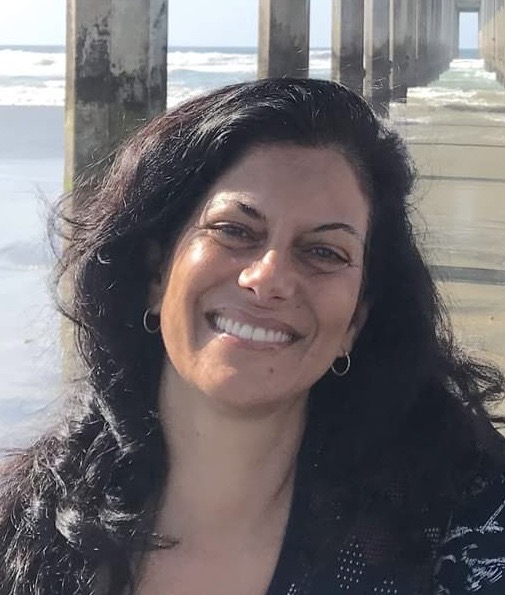
Director, Biophotonics Core
dboassa@salk.edu
Dr. Daniela Boassa completed her Ph.D. in Neuroscience at the University of Arizona, Tucson. In 2005 she joined the National Center for Microscopy and Imaging Research at UCSD where she led activities related to the development, characterization, enhancement, and dissemination of novel molecular-genetic probes and chemical-labeling approaches for state-of-the-art correlated light and electron microscopy (CLEM) and their application to biomedical research. She has expertise in neuroscience, cell and molecular biology and sophisticated microscopy and analysis. Over the years, she trained and mentored local, national, and international research groups on CLEM technologies. She takes pride in the success and achievements of the students and young scientists she mentored over the years. Dr. Boassa joined the Salk Institute in 2024.
List of publications: https://scholar.google.com/citations?user=ztNqcs8AAAAJ&hl=en

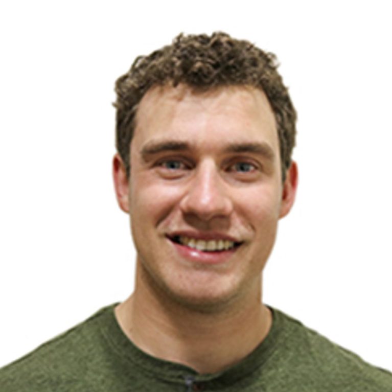
Senior Imaging and Microscopy Specialist
sweisernovak@salk.edu
After completing his B.S. in Biomedical Toxicology at the University of Guelph in Ontario, Canada, Sammy Weiser Novak moved to the University of Victoria to perform his M.S. in the laboratory of Dr. Patrick Nahirney, the author of Netter’s Histology. There Sammy used electron microscopy and computational image analysis to study the ultrastructure and morphology of hippocampal synapses in a mouse model of fragile X syndrome. Afterwards, Sammy worked in Dr. Joel Kubby’s lab at UC-Santa Cruz developing a 2-photon microscope with adaptive optics, gaining valuable hands-on experience with custom hardware and advanced light microscope systems. He then joined the laboratory of Dr. Marie-Eve Trembley to establish 3D electron microscopy using a new FIB-SEM system recently acquired for her lab. Sammy’s current research interests include synapse structure and function, 3D electron microscopy techniques including correlative and cryo-electron microscopy, and machine-learning assisted image processing, analysis, and segmentation.

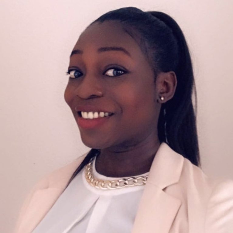
Light Microscopy Specialist
equansah@salk.edu
Elsie obtained her Bachelor degree in Physics from the University of Cape Coast, Ghana. After, she pursued her Master degree in Photonics from the Friedrich Schiller University in Jena, Germany, which she completed in 2018. She continued her academic pursuit by undertaking a Ph.D. at the Leibniz Institute for Photonic Technology in Germany.
With years of experience as a research scientist in the field of spectroscopy and microscopy, she is an expert in non-linear multimodal imaging, including coherent anti-Stokes Raman scattering (CARS), two-photon excited fluorescence (TPEF), and second harmonic generation (SHG). During her doctoral studies, she specialized in the optimization of label-free imaging modalities, leveraging these advanced techniques for the early detection of cancerous cells and tissues.
She worked on the automatic detection of breast cancer using nonlinear imaging methods, revolutionizing the way clinicians diagnose and monitor this prevalent disease. Additionally, she conducted research on intestinal epithelial barrier integrity in patients with ulcerative colitis, employing label-free techniques to gain insights into the morpho-chemical changes in disease pathology. The culmination of this research set the stage for the development of a fiber-based CARS/TPEF/SHG imaging setup for an endomicroscopic imaging probe, which holds the potential to revolutionize medical diagnostics by providing clinicians with real-time imaging capabilities directly within hospitals.