May 7 - 10, 2018

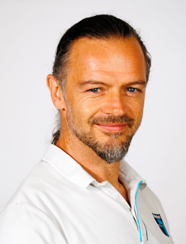
JB Sibarita has a PhD thesis in Physics and is expert in live cell microscopy and image analysis. He is heading a CNRS R&D team “Quantitative Imaging of the Cell” at the Interdisciplinary Institute for Neuroscience in Bordeaux with a strong emphasis on quantitative super-resolution microscopy to decipher protein organization and dynamics in well-controlled cellular environments. He is authors of more than 60 scientific peer-review articles, 3 patents and 5 industrial technology transfer.
http://www.iins.u-bordeaux.fr/TEAM-SIBARITA

Eric Hosy has a PhD cellular physiology and is expert neuroscience more particularly on the use of electrophysiology and super-resolution imaging to understand synaptic transmission. He is PI at the CNRS in the group of Daniel Choquet at the Interdisciplinary Institute for Neuroscience in Bordeaux. He tightly works in collaboration with the Sibarita’s group to develop and transfer super-resolution techniques to other models (plant, bacteria, etc.). He signed 20 papers the last 5 years in journal as Nature, nature methods and neurons.
http://www.iins.u-bordeaux.fr/Receptor-Organization-and-Synaptic-189

As Director of the Advanced Biophotonics Core Facility at the Salk Institute, Dr. Manor’s primary focus is the integration and application of optical and charged particle detection technologies to study problems of critical biological significance. He is a cellular and molecular biophysicist by doctoral training. During his Ph.D. and postdoctoral training he used advanced light and electron microscopy as computational methods to determine how the cellular cytoskeleton regulates organelle size, shape, and dynamics. His graduate work with Dr. Bechara Kachar (NIH), and his postdoctoral training with Dr. Jennifer Lippincott-Schwartz (NIH and Janelia Farms) provided Dr. Manor with a broad range of training in addressing key biological questions using advanced imaging approaches, such as superresolution and live cell imaging, automated analysis and segmentation of microscopy data, and computational modeling of biophysical and biochemical dynamics in the cell. By the time Dr. Manor completed his postdoctoral training, he had published 17 peer-reviewed publications, all of which relied on Dr. Manor’s imaging or image analysis skills. Dr. Manor’s main biological interests are actin-dependent organelle dynamics, inner ear hair cell stereocilia, and neuronal synapse structure and function.