
August 10, 2020
Molecules of actin help control the division of mitochondria, with implications for disease
Molecules of actin help control the division of mitochondria, with implications for disease
LA JOLLA—While your skeleton helps your body to move, fine skeleton-like filaments within your cells likewise help cellular structures to move. Now, Salk researchers have developed a new imaging method that lets them monitor a small subset of these filaments, called actin.
“Actin is the most abundant protein in the cell, so when you image it, it’s all over the cell,” says 尤里·马诺尔, director of Salk’s Biophotonics Core facility and corresponding author of the paper. “Until now, it’s been really hard to tell where individual actin molecules of interest are, because it’s difficult to separate the relevant signal from all the background.”
点击此处 用于高分辨率图像。.
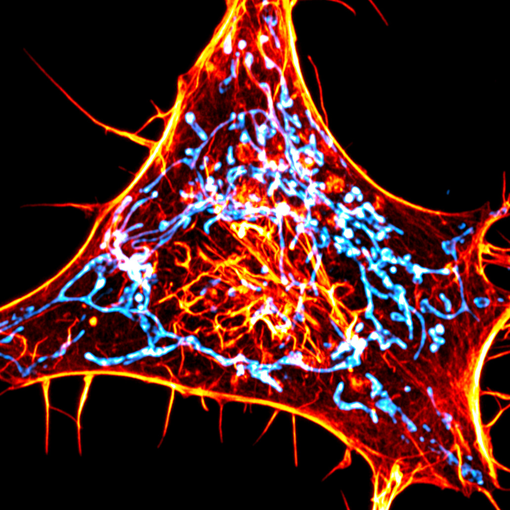
Credit: Salk Institute/Waitt Advanced Biophotonics Center
With the new imaging technique, the Salk team has been able to home in on how actin mediates an important function: helping the cellular “power stations” known as mitochondria divide in two. The work, which appeared in the journal 自然方法 on August 10, 2020, could provide a better understanding of mitochondrial dysfunction, which has been linked to cancer, aging, and neurodegenerative diseases.
Mitochondrial fission is the process by which these energy-generating structures, or organelles, divide and multiply as part of normal cellular maintenance; the organelles divide not only when a cell itself is dividing, but also when cells are under high amounts of stress or mitochondria are damaged. However, the exact way in which one mitochondrion pinches off into two mitochondria has been poorly understood, particularly how the initial constriction happens. Studies have found that removing actin from a cell entirely, among many other effects, leads to less mitochondrial fission, suggesting a role for actin in the process. But destroying all the actin causes so many cellular defects that it’s hard to study the protein’s exact role in any one process, the researchers say.
So, Manor and his colleagues developed a new way to image actin. Rather than tag all the actin in the cell with fluorescence, they created an actin probe targeted to the outer membrane of mitochondria. Only when actin is within 10 nanometers of the mitochondria does it attach to the sensor, causing the fluorescence signal to increase.
Rather than see actin scattered haphazardly around all mitochondrial membranes, as they might if there were no discrete interactions between actin and the organelles, Manor’s team saw bright hotspots of actin. And when they looked closely, the hotspots were located at the same locations where another organelle called the endoplasmic reticulum crosses the mitochondria, previously found to be fission sites. Indeed, as the team watched actin hotspots light up and disappear over time, they discovered that 97 percent of mitochondrial fission sites had actin fluorescing around them. (They speculate that there was also actin at the other 3 percent of fission sites, but that it wasn’t visible).
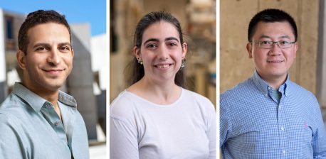
“This is the clearest evidence I’ve ever seen that actin is accumulating at fission sites,” says Cara Schiavon, co-first author of the paper and a joint postdoctoral fellow in the labs of Uri Manor and Salk Professor 杰拉尔德·沙德尔. “It’s much easier to see than when you use any other actin marker.”
By altering the actin probe so that it attached to the endoplasmic reticulum membrane rather than the mitochondria, the researchers were able to piece together the order in which different components join the mitochondrial fission process. The team’s results suggest that the actin attaches to the mitochondria before it reaches the endoplasmic reticulum. This lends important insight towards how the endoplasmic reticulum and mitochondria work together to coordinate mitochondrial fission.
点击此处 用于高分辨率图像。.
版权:萨克研究所
In additional experiments described in a pre-print manuscript available on bioRxiv, Manor’s team also reports that the same accumulation of endoplasmic reticulum-associated actin is seen at the sites where other cellular organelles—including endosomes, lysosomes and peroxisomes—divide. This suggests a broad new role for a subset of actin in organelle dynamics and homeostasis (physiological equilibrium).
In the future, the team hopes to look at how genetic mutations known to alter mitochondrial dynamics might also affect actin’s interactions with the mitochondria. They also plan to adapt the actin probes to visualize actin that’s close to other cellular membranes.
“This is a universal tool that can now be used for many different applications,” says Tong Zhang, a light microscopy specialist at Salk and co-first author of the paper. “By switching out the targeting sequence or the nanobody, you can address other fundamental questions in cell biology.”
“We’re in a golden age of microscopy, where new instruments with ever higher resolution are always being invented; but in spite of that there are still major limitations to what you can see,” says Manor. “I think combining these powerful microscopes with new methods that select for exactly what you want to see is the next generation of imaging.”
Other researchers on the study were Pauline Wales, Leonardo Andrade, Melissa Wu, Tsung-Chang Sung, Yelena Dayn, and Gerald Shadel of Salk; Bing Zhao and Robert Grosse of the University of Freiburg; Andrew Moore of the Howard Hughes Medical Institute; and Jasmine Feng and Omar Quintero of the University of Richmond.
The Waitt Advanced Biophotonics Center (home of Salk’s Biophotonics Core facility) is funded by the Waitt Foundation and National Cancer Institute. The work and researchers involved were also supported by the Salk Transgenic Core Facility, National Institutes of Health, National Institute of General Medical Sciences, Human Frontier Science Program, Centre for Integrative Biological Signaling Studies, and the University of Richmond School of Arts & Sciences.
DOI: 10.1038/s41592-020-0926-5
日记
自然方法
作者
Cara R. Schiavon, Tong Zhang, Bing Zhao, Andrew S. Moore, Pauline Wales, Leonardo Andrade, Melissa Wu, Tsung-Chang Sung, Yelena Dayn, Jasmine W. Feng, Omar A. Quintero, Gerald S. Shadel, Robert Grosse, and Uri Manor
宣传办公室
电话:(858) 453-4100
press@salk.edu
萨尔克研究所是一个独立的非营利性研究机构,由首个安全有效的脊髓灰质炎疫苗的研发者乔纳斯·索尔克于1960年创立。该研究所的使命是推动以合作、敢于冒险为特点的基础性研究,以应对癌症、阿尔茨海默病和农业脆弱性等社会最紧迫的挑战。这项基础科学支撑着所有的转化研究,产生有助于全球新药和创新的见解。.


