
July 11, 2013
Salk researchers' findings on chromosome shortening suggest a potential target to arrest cancer cell growth
Salk researchers' findings on chromosome shortening suggest a potential target to arrest cancer cell growth
LA JOLLA,CA—A team of scientists at the Salk Institute for Biological Studies has identified why disruption of a vital pathway in cell cycle control leads to the proliferation of cancer cells. Their findings on telomeres, the stretches of DNA at the ends of chromosomes that protect our genetic code and make it possible for cells to divide, suggest a potential target for preventive measures against cancer, aging and other diseases. The findings were published July 11, 2013 in Molecular Cell.
Telomeres have been compared to the plastic tips at the end of shoelaces because they prevent the ends of chromosomes from fraying and sticking to each other, which scrambles the genetic information and may promote cancer. They are crucial to DNA replication, tumor suppression and aging. Each time a human cell divides, its telomeres become shorter. When they become too short, the cell can no longer divide and becomes inactive, or “senescent,” or dies. Cells can escape this fate by activating an enzyme called telomerase, which prevents telomeres from getting shorter and allows the cells to continue to grow and divide. Uncontrolled cellular growth is a primary hallmark of cancer cells, and shortened telomeres have been identified in pancreatic, bone, prostate, bladder, lung, kidney and head and neck cancers.
“As telomeres shorten during normal [cellular] aging, they activate a DNA damage response to arrest cell growth, which protects our DNA from harm,” says senior study author 扬·卡尔塞德, ,索尔克研究所的教授 分子与细胞生物学实验室 and holder of the Donald and Darlene Shiley Chair.
Karlseder and his team identified that cell growth arrest due to shortening telomeres is confined to one specific portion of the cell cycle, called the G1 phase, which is the most protected stage of the cell cycle. “The pathway controlling G1-phase growth arrest, however, is commonly altered in cancer cells, allowing cancer cells to divide despite shortened telomeres, which can lead to the genomic instability seen in malignant cells.”
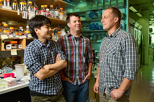
From left: Salk scientists Makoto Hayashi, Anthony Cesare and Jan Karlseder
图片:由萨克生物研究所提供
In the study, Karlseder and his colleagues mimicked the process of cellular aging by partially removing a protein called TRF2 from the telomeres of human fibrosarcoma (a type of cancer that affects connective tissue) cells. By doing so, they were able to experimentally reproduce the process that occurs naturally as cells age. This telomere “deprotection” exposed the ends of chromosomes during certain stages of the cell cycle. In this state, they found that telomeres exhibited a partial DNA damage response: the ends of chromosomes were protected against fusing and fraying, but cell growth was still arrested.
“Basically,” says lead author Anthony Cesare, a research associate in Karlseder’s laboratory, “there’s cell growth arrest without genomic instability. Thus, telomere aging, in normal, healthy cells and living organisms, means cell arrest, but no harmful genetic effects.”
The Salk scientists identified the p53 pathway, a molecular mechanism that normally protects a cell’s genetic material and suppresses tumors, as the key player in the response to telomere deprotection. When cells lose the function of p53, the gene at the center of the pathway, they can no longer arrest cells in the G1 phase, an important point in the cell cycle for repairing DNA damage or, if the damage cannot be repaired, targeting the cell for programmed death. Most commonly, p53 is lost in cancer cells due to a mutation in the p53 gene or the inactivation of p53 protein function through infection from cancer-causing viruses.
Because telomere deprotection results in a partial DNA damage response that only arrests cells in G1 through the p53 pathway, once cells lose p53 function telomere deprotection no longer arrests growth. “Cells without functional p53 are able to divide with deprotected telomeres, which causes genomic instability, a common feature of malignant cells,” says Karlseder.
Karlseder and his colleagues believe that better understanding the telomere shortening process may lead to the ability to influence cellular aging and, as a result, stunt cancer cell growth. They say the next step is determining why this deprotection response is muted in cancer cells, and possibly affecting this process to prevent cancer cells from growing.
Other researchers on the study were Makoto T. Hayashi and Laure Crabbe of the Salk Institute. The work was supported by the 美国国立卫生研究院, the John Sabo Trust, the Highland Street Foundation, , 那个 Human Frontier Science Program, , 那个 Japan Society for the Promotion of Science and the Salk Institute Glenn Center for Aging.
关于索尔克生物研究所:
索尔克生物研究所是世界顶尖的基础研究机构之一,其国际知名的教职人员在一个独特、协作和富有创造性的环境中,深入探究生命科学的基本问题。索尔克科学家们致力于发现和指导未来几代研究人员,通过研究神经科学、遗传学、细胞和植物生物学以及相关学科,在癌症、衰老、阿尔茨海默氏症、糖尿病和传染病的认识方面做出了开创性的贡献。.
学院取得了许多成就,获得了包括诺贝尔奖和美国国家科学院院士在内的无数荣誉。该研究所由脊髓灰质炎疫苗先驱 Jonas Salk 博士于 1960 年创立,是一家独立的非营利组织和建筑地标。.
日记
Molecular Cell
作者
Anthony J. Cesare, Makoto T. Hayashi, Laure Crabbe, and Jan Karlseder
宣传办公室
电话:(858) 453-4100
press@salk.edu


