
February 10, 2012
Discovery provides clues to development of neurological diseases and cancer
Discovery provides clues to development of neurological diseases and cancer
LA JOLLA, CA—Researchers at the Salk Institute have discovered a startling feature of early brain development that helps to explain how complex neuron wiring patterns are programmed using just a handful of critical genes. The findings, published in 细胞, may help scientists develop new therapies for neurological disorders, such as amyotrophic lateral sclerosis (ALS), and provide insight into certain cancers.
The Salk researchers discovered that only a few proteins on the leading edge of a motor neuron’s axon – its outgoing electrical “wire” – and within the extracellular soup it travels through guide the nerve as it emerges from the spinal cord. These molecules can attract or repel the axon, depending on the long and winding path it must take to finally connect with its target muscle.
“The budding neuron has to detect the local environment it is growing through and decide where it is, and whether to grow straight, move to the left or right, or stop,” says the study’s senior investigator, 萨姆·普法夫, ,索尔克研究所的教授 基因表达实验室 and a Howard Hughes Medical Institute investigator.
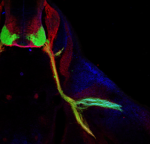
This image shows nerves (labeled in green) that control body movements emerging from the spinal cord of a mouse (upper left, in cross section) and connecting to muscles in the base of the leg.
The Salk researchers discovered that a combination of genes direct nerves to split in the leg (lower right) to make the proper connections with their target muscles during early development.
Image: Courtesy Dario Bonanomi, Salk Institute for Biological Studies
“It does this by mixing and matching just a handful of protein products to create complexes that tell a growing neuron which way to go, in the same way that a car uses the GPS signals it receives to guide it through an unfamiliar city,” he says.
The brain contains millions of times the number of neuron connections than the number of genes found in the DNA of brain cells. This is one of the first studies to try and understand how a growing neuron integrates many different pieces of information in order to navigate to its eventual target and make a functional connection.
“We focused on motor neurons that control muscle movements, but the same kind of thing is going on throughout embryonic development of the entire nervous system, during which millions of axons make trillions of decisions as they move to their targets,” he says. “It is the exquisite specificity with which they grow that underlies the basic architecture and proper function of the nervous system.”
These findings might eventually shed new light on a number of clinical disorders related to faulty nerve cell functioning, such as ALS, which is also known as Lou Gehrig’s disease, says the first author on the paper, Dario Bonanomi, a post-doctoral researcher in Pfaff’s laboratory.
“These are the motor neurons that die in diseases like Lou Gehrig’s disease and that are linked to a genetic disorder in children known as spinal muscle atrophy,” Bonanomi says.
“It is also a jumping off point to try and understand the basis for defects that might arise during fetal development of the nervous system,” he added. “A better understanding of those signals might help to be able to regenerate and rewire circuits following diseases or injuries of the nervous system.”
The researchers say the study also offers insights into cancer development, because a protein the researchers found to be crucial to the “push and pull” signaling system – Ret- is also linked to cancer. Mutations that activate Ret are linked to a number of different kinds of tumors.
The other protein receptors described in the study, known as Ephs, have also been implicated in cancer, Pfaff says.
“This study suggests that the way cells detect signals in their environment is likely a universal strategy,” he says, “and we know that genes and proteins known to function primarily during embryonic development have been linked to cancer.”
“Controlling neuronal growth requires very potent signaling molecules, and it makes sense they would be linked to disease,” Pfaff says. “We hope our findings help further unravel these connections.”
The study was funded by the National Institute of Neurological Disorders and Stroke and by the 霍华德·休斯医学研究所.
Co-authors include, from Salk, Onanong Chivatakar, Ge Bai, and Karen Lettieri; Houari Abdesselem and Brian A. Pierchala, from the University of Michigan School of Dentistry; and Till Marquardt, from the European Neuroscience Institute-Göttingen, in Germany.
关于索尔克生物研究所:
索尔克生物研究所是世界顶尖的基础研究机构之一,其国际知名的教职人员在一个独特、协作和富有创造性的环境中,深入探究生命科学的基本问题。索尔克科学家们致力于发现和指导未来几代研究人员,通过研究神经科学、遗传学、细胞和植物生物学以及相关学科,在癌症、衰老、阿尔茨海默氏症、糖尿病和传染病的认识方面做出了开创性的贡献。.
学院取得了许多成就,获得了包括诺贝尔奖和美国国家科学院院士在内的无数荣誉。该研究所由脊髓灰质炎疫苗先驱 Jonas Salk 博士于 1960 年创立,是一家独立的非营利组织和建筑地标。.
日记
细胞
标题
Ret Is a Multifunctional Coreceptor that Integrates Diffusible- and Contact-Axon Guidance Signals
作者
Dario Bonanomi, Onanong Chivatakarn, Ge Bai, Houari Abdesselem, Karen Lettieri, Till Marquardt, Brian A. Pierchala and Samuel L. Pfaff
宣传办公室
电话:(858) 453-4100
press@salk.edu


