
2016年4月28日
Salk imaging technologies offer new window into spinal cord to understand touch and pain sensations
Salk imaging technologies offer new window into spinal cord to understand touch and pain sensations
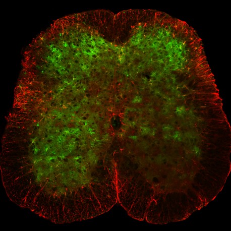
LA JOLLA—A microscope about the size of a penny is giving scientists a new window into the everyday activity of cells within the spinal cord. The innovative technology revealed that astrocytes—cells in the nervous system that do not conduct electrical signals and were traditionally viewed as merely supportive—unexpectedly react to intense sensation.
The new miniaturized microscope and related imaging methods, described by Salk Institute scientists on April 28, 2016 in Nature Communications, offer unprecedented insight into nervous system function and could lead to novel pain treatments for spinal cord injuries, chronic itch and neurodegenerative diseases such as amyotrophic lateral sclerosis (ALS).
点击此处 用于高分辨率图像。.
版权:萨克研究所
The spinal cord is crucial for sensing and responding to the world. Sometimes it even works independently from the brain, such as when your hand recoils from a hot stove before the sensation has fully registered. But it is unknown exactly how the cells within the spinal cord encode these and other feelings from the skin or internal organs.
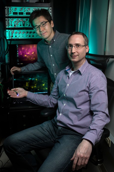
In the new study, senior author 阿克塞尔·尼默雅恩, an assistant professor in Salk’s 韦特先进生物光子学中心, and his team improved upon the miniaturized microscopes they first described back in 2008. The researchers’ new version—which features numerous hardware and software improvements—enabled them to visualize changes in cellular activity in awake, roaming mice.
“For a long time, researchers have dreamed of being able to record cellular activity patterns in the spinal cord of an awake animal. On top of that, we can now do this in a freely behaving animal, which is very exciting,” says first author Kohei Sekiguchi, a Salk researcher and PhD student at the 加州大学圣地亚哥分校.
Most of the Salk team’s previous work focused on deploying microscopes to observe the brains of living animals. The spinal cord, by contrast, presented a bigger challenge for several reasons. For example, unlike the brain, multiple, independently moving vertebrae surround the spinal cord. The spinal cord is also closer to pulsating organs (heart and lungs), which can hinder stable views of the cells within. However, by developing new microscopy and procedural and computational approaches, the team was able to overcome these challenges and capture the action of living cells in real time and during vigorous movements.
Click here for a high-resolution image
版权:萨克研究所
In the new work, the group found that distinct stimuli—such as light touch or pressure—activate different subsets of spinal sensory neurons. They also found that certain features, like the intensity or duration of a given stimulus, are reflected in the activity of the neurons.
To the team’s surprise, astrocytes, traditionally thought to be passive support cells, also respond to stimuli (albeit differently than the neurons). Though the astrocytes cannot send electrical signals like neurons can, they generated their own chemical signals in a coordinated way during intense stimuli.
Nimmerjahn is excited about this result because his group has a longstanding interest in understanding astrocytes and their roles in nervous system function and disease. These cells are increasingly appreciated as important players in how the nervous system develops and operates and could serve as promising new drug targets, he says.
“Not only can we now study normal sensory processing, but we can also look at disease contexts like spinal cord injury and how treatments actually affect the cells,” says Nimmerjahn.
The team is now working to simultaneously record touch or pain-related activity in the brain and spinal cord using additional iterations of the miniaturized microscopes, which allow them to monitor and manipulate multiple cell types at even higher resolutions.
Other researchers on the paper include the Salk Institute’s Pavel Shekhtmeyster, Katharina Merten, Alexander Arena, Daniela Cook, Elizabeth Hoffman and Alexander Ngo.
该项工作获得了以下机构的资助: 美国国立卫生研究院, , 那个 丽塔·艾伦基金会, Whitehall Foundation 和 Brain Research Foundation; funds from the Waitt Foundation, Hearst Foundations and the Richard Allan Barry Family Charitable Foundation; and research fellowships from the Nakajima Foundation, Mary K. Chapman Foundation, Jesse and Caryl Philips Foundation, the 罗斯希尔斯基金会, Deutsche Forschungsgemeinschaft (DFG) and the Catharina Foundation.
日记
Nature Communications
作者
Kohei J. Sekiguchi, Pavel Shekhtmeyster, Katharina Merten, Alexander Arena, Daniela Cook, Elizabeth Hoffman, Alexander Ngo, and Axel Nimmerjahn
宣传办公室
电话:(858) 453-4100
press@salk.edu
萨尔克研究所是一个独立的非营利性研究机构,由首个安全有效的脊髓灰质炎疫苗的研发者乔纳斯·索尔克于1960年创立。该研究所的使命是推动以合作、敢于冒险为特点的基础性研究,以应对癌症、阿尔茨海默病和农业脆弱性等社会最紧迫的挑战。这项基础科学支撑着所有的转化研究,产生有助于全球新药和创新的见解。.


