
June 4, 2026
Salk Institute scientists use new 3D reconstructions and computer simulations to reveal previously unmeasurable changes in synaptic structure during key learning process in brain, opening new avenues for understanding neurological disease
LA JOLLA—Inside the brain is a dense network of neurons that receive, process, and relay information. The synapse, where neurons meet, is the epicenter of this communication. Neurons that send information, called presynaptic neurons, hold tiny packages of neurotransmitters—waiting for a chemical signal from the brain to be released. How this system is regulated by the brain during periods of learning has, until now, been out of reach.
Salk Institute scientists have finally grasped an answer, discovering that the density of synaptic vesicles, where neurotransmitters are held in the presynaptic terminal, is actively regulated by the brain during long-term potentiation (LTP). LTP is a neuronal process widely regarded as key to learning and memory. These findings reveal that synaptic vesicle density is dynamic and regulated by the brain during LTP.
该研究发表在 美国国家科学院院刊 on May 26, 2026, lays the groundwork for understanding how synaptic vesicle density and its regulation may contribute to aging and neurological disease.
“Altering synapse strength is essential for learning, as it allows neural circuits to adapt to environmental changes—we want to know what exact structural and functional changes are happening,” says senior author 特伦斯·塞津斯基博士, a professor and Francis Crick Chair at Salk. “Uncovering the molecular mechanisms underlying synaptic vesicle clustering is fundamental to understanding synaptic transmission, learning, and memory.”
To discover that the brain regulates vesicle density, the team developed methods to generate 3D reconstructions from electron microscopy images and to quantify synapse structure.
3D reconstruction and computer simulations enabled researchers to link vesicle density to the viscosity of synaptic vesicles in the cluster. This technical breakthrough is the heart of the research—with 3D reconstructions and computer simulations, the scientists could observe and quantify changes within the synapse that were previously impossible to study.
“Once you have observed something and know how to measure something that no one has been able to measure before, this lets you look at lots of things in a new way,” says coauthor of the study Thomas Bartol, PhD, a staff researcher in Sejnowski’s lab.
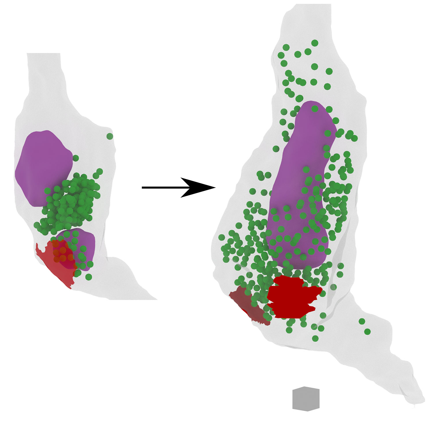
The team examined mammalian hippocampi to uncover changes in synaptic vesicle density under controlled conditions before and after inducing LTP. The neurons were then visualized with their novel 3D reconstruction methods from electron microscopy, and their structure was quantified.
The major discovery was that vesicle cluster density was not static nor routine; rather, it shifted in response to LTP. This means that the brain is deliberately regulating the neuronal network at the level of individual synapses. As the neurons learned, vesicle density decreased compared to control neurons that were not learning. This reduction in density was found, through computer simulations, to be associated with an increase in synaptic vesicle mobility.
Previous studies of synaptic plasticity have found many structural changes associated with the strengthening and weakening of synapses. The new findings provide novel insight into how synapse strength changes after LTP.
The implications of this discovery stretch far beyond basic learning and memory. Synaptic dysfunction is thought to be the cause of a wide range of neurological diseases and age-related neurodegeneration. However, scientists have lacked the tools necessary to pinpoint what goes wrong on the structural level.
“We are showing that neuron properties change during LTP, but this could also happen in other contexts, like aging. I definitely think that it’s a very exciting area of research,” says first author of the study, Guadalupe Garcia, PhD, a postdoctoral researcher in Sejnowski’s lab.
“We hope to investigate these same processes in young and adult models, to see if and how synaptic vesicle alterations contribute to age-associated diseases like Alzheimer’s,” adds Sejnowski.
With this new visualization technology, new opportunities for quantitative analysis emerge. Understanding how the regulation of synaptic vesicle clusters differs between healthy individuals and those with aging-related or neurological conditions could help researchers pinpoint the specific mechanisms driving disease—and potentially point toward new therapeutic targets.
Other authors include Priyal Badala of Salk, as well as Lyndsey Kirk and Kristen Harris of University of Texas at Austin.
The work was supported by the National Science Foundation (1707356, 2014862) and National Institutes of Health (R01MH095980, R56MH139176).
DOI: 10.1073/pnas.2522754123
日记
美国国家科学院院刊
作者
Guadalupe C. Garcia, Thomas M. Bartol, Lyndsey M. Kirk, Priyal Badala, Kristen M. Harris, Terrence J. Sejnowski
宣传办公室
电话:(858) 453-4100
press@salk.edu
萨尔克研究所是一个独立的非营利性研究机构,由首个安全有效的脊髓灰质炎疫苗的研发者乔纳斯·索尔克于1960年创立。该研究所的使命是推动以合作、敢于冒险为特点的基础性研究,以应对癌症、阿尔茨海默病和农业脆弱性等社会最紧迫的挑战。这项基础科学支撑着所有的转化研究,产生有助于全球新药和创新的见解。.


