
May 2, 2016
Salk study is first to closely follow development of new neurons in the adult brain, giving potential insight into neurodevelopmental disorders such as autism and schizophrenia
Salk study is first to closely follow development of new neurons in the adult brain, giving potential insight into neurodevelopmental disorders such as autism and schizophrenia
LA JOLLA—When tweaking its architecture, the adult brain works like a sculptor—starting with more than it needs so it can carve away the excess to achieve the perfect design. That’s the conclusion of a new study that tracked developing cells in an adult mouse brain in real time.
点击此处 用于高分辨率图像
版权:萨克研究所
New brain cells began with a period of overgrowth, sending out a plethora of neuronal branches, before the brain pruned back the connections. The observation, described May 2, 2016 in 自然-神经科学, suggests that new cells in the adult brain have more in common with those in the embryonic brain than scientists previously thought and could have implications for understanding diseases including autism, intellectual disabilities and schizophrenia.
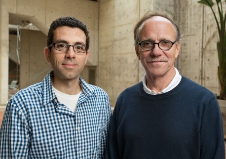
“We were surprised by the extent of the pruning we saw,” says senior author 鲁斯蒂·盖奇, a professor in Salk’s Laboratory of Genetics and holder of the Vi and John Adler Chair for Research on Age-Related Neurodegenerative Disease.
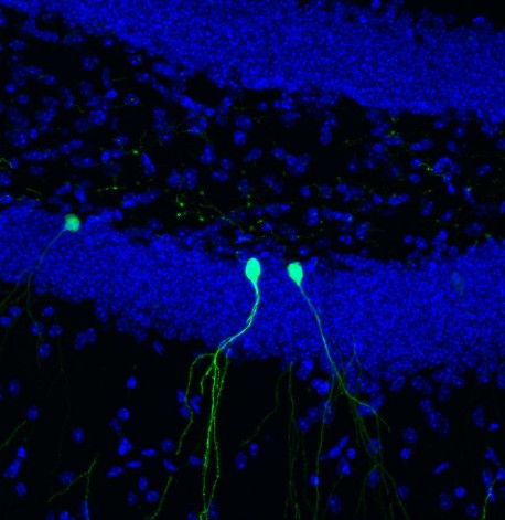
While most of the brain’s billions of cells are formed before birth, Gage and others previously showed that in a few select areas of the mammalian brain, stem cells develop into new neurons during adulthood. In the new study, Gage’s group focused on cells in the dentate gyrus, an area deep in the brain thought to be responsible for the formation of new memories. The scientists used a new microscopy technique to observe new cells being formed in the dentate gyrus of adult mice.
“This is the first time we’ve been able to image dentate neurons growing in a living animal,” says Tiago Gonçalves, a research associate in the Gage lab and first author of the new paper.
点击此处 用于高分辨率图像
版权:萨克研究所
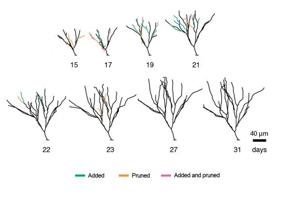
Gonçalves and Gage followed—on a daily basis—the growth of neurons over several weeks. When animal were housed in environments with lots of stimuli—running wheels, plastic tubes, and domes—the new cells grew quickly, sending out dozens of branches called dendrites which receive electrical signals from surrounding neurons. When kept in empty housing, the new neurons grew slightly slower and sent out, on average, a few less dendrites. But, in both cases, the dendrites of the new cells began to be pruned back.
“What was really surprising was that the cells that initially grew faster and became bigger were pruned back so that, in the end, they resembled all the other cells,” says Gonçalves. He and his colleagues went on to show that changing signaling pathways could mimic some of the effects of the complex environment—cells grew more initially, but also pruned back earlier.
点击此处 用于高分辨率图像
版权:萨克研究所
So why would the brain spend energy developing more dendrites than needed? The researchers suspect that the more dendrites a neuron starts with, the more flexibility it has to prune back exactly the right branches.
“The results suggest that there is significant biological pressure to maintain or retain the dendrite tree of these neurons,” says Gage.
Defects in the dendrites of neurons have been linked to numerous brain disorders including schizophrenia, Alzheimer’s, epilepsy and autism. Charting how the brain shapes these branches—both during embryonic development and in adulthood—may be the key to understanding mental health.
“This also has big repercussions for regenerative medicine,” says Gonçalves. “Could we replace cells in this area of the brain with new stem cells and would they develop in the same way? We don’t know yet.”
Other researchers on the study were Cooper W. Bloyd, Matthew Shtrahman, Stephen T. Johnston, Simon T. Schafer, Sarah L. Parylak, Tranh Tran, and Tina Chang of the Salk Institute.
The work and the researchers involved were supported by grants from The James S. McDonnell Foundation, CIRM, G. Harold & Leila Y. Mathers Charitable Foundation, Annette Merle-Smith, JBP Foundation, 美国国立卫生研究院 和 利昂娜·M·哈里·B·赫尔姆斯利慈善信托.
日记
自然-神经科学
作者
J. Tiago Gonçalves, Cooper W. Bloyd, Matthew Shtrahman, Stephen T. Johnston, Simon T. Schafer, Sarah L. Parylak, Tranh Tran, Tina Chang, and Fred H. Gage of the Salk Institute
宣传办公室
电话:(858) 453-4100
press@salk.edu
萨尔克研究所是一个独立的非营利性研究机构,由首个安全有效的脊髓灰质炎疫苗的研发者乔纳斯·索尔克于1960年创立。该研究所的使命是推动以合作、敢于冒险为特点的基础性研究,以应对癌症、阿尔茨海默病和农业脆弱性等社会最紧迫的挑战。这项基础科学支撑着所有的转化研究,产生有助于全球新药和创新的见解。.


