
April 23, 2026
Salk Institute scientists find correcting dysregulations in star-shaped brain cells called astrocytes improves some fragile X syndrome symptoms, making the cells potential future therapeutic targets
Salk Institute scientists find correcting dysregulations in star-shaped brain cells called astrocytes improves some fragile X syndrome symptoms, making the cells potential future therapeutic targets
LA JOLLA—Fragile X syndrome (FXS) is an inherited genetic developmental condition that strongly impacts brain development. Despite the syndrome stemming from altered genetic code for the single protein fragile X messenger ribonucleoprotein (FMRP), its symptoms are broad and variable; people with FXS can have a range of behavioral and physical symptoms, and around 40 percent of people with FXS also have autism spectrum disorder. There is currently no cure for FXS; treatments are limited to medications and therapies to help manage symptoms.

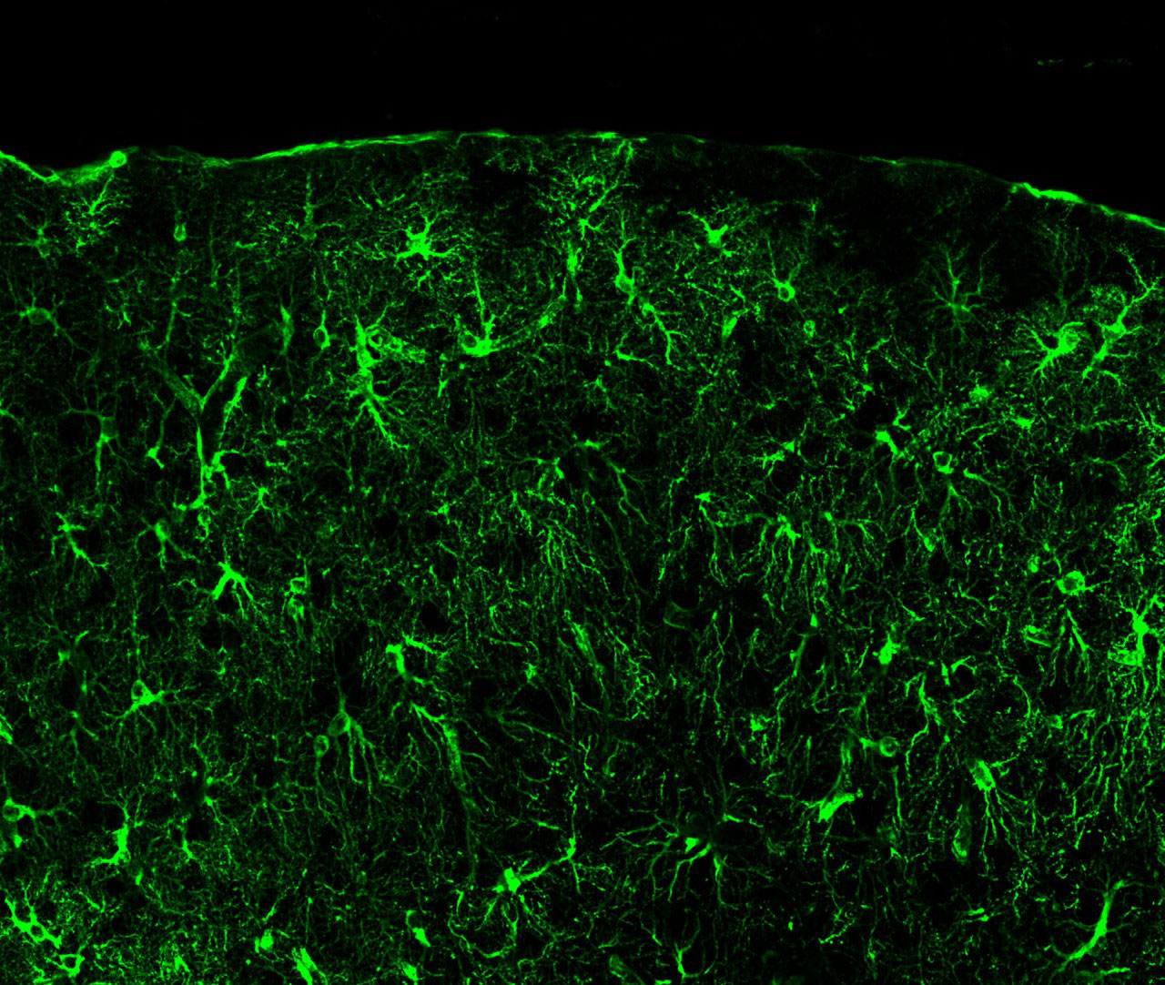
New research from the Salk Institute found how star-shaped brain cells called astrocytes contribute to some FXS symptoms. What’s more, they found that a protein pathway commonly upregulated in FXS astrocytes could be suppressed to lessen those symptoms—meaning less severe seizures and restored molecular balances in a mouse model of FXS.
Die Ergebnisse, veröffentlicht in Nature Communications on April 23, 2026, validate the importance of studying astrocytes in FXS research and are a promising step toward future therapeutics for FXS and other developmental conditions, like Down syndrome or Rett syndrome.
“This dataset identifies astrocyte-specific alterations to proteins that allow astrocytes to regulate neurons in a whole-brain context,” says senior author of the study Nicola Allen, PhD, professor and Roger Guilleman Chair at Salk. “It’s a great resource for fragile X syndrome researchers, but also for the scientific community beyond any single disorder or condition. Using this approach, we can study astrocyte protein changes within a whole brain context and make their inclusion easier moving forward.”
FXS is the most common cause of inherited intellectual disability, so understanding exactly how it manifests in the brain is crucial. It makes sense, then, that scientists have been studying the role neurons play in FXS.
One consistent finding has been dysfunctional synapses, which are the junctions between neurons where information exchange occurs. Research has shown that in FXS, there are structural differences in neurons’ dendritic spines, the site of input during synaptic information exchange.
These two dysfunctions have something in common: Both synapse activity and dendritic spine morphology are regulated by astrocytes. Astrocytes are abundant non-neuronal glial cells found throughout the brain and are crucial for the development and maintenance of healthy neurons.
This link between neurons and astrocytes in FXS is no surprise to the astrocyte-savvy researchers in Allen’s lab.
“Recent research, including in our lab, has shown that astrocytes have many changed genes and proteins in fragile X syndrome,” says first author James Deng, who led this project as a graduate student researcher in Allen’s lab. “Our study accelerates this ongoing work by studying fragile X syndrome astrocytes through multiple angles in a living system, which gives us novel insights into those changes.”
Building off their previous findings that genes and proteins are dysregulated in FXS astrocytes when isolated and grown in a dish, the Salk team zeroed in on one specific dysregulated pathway: bone morphogenetic protein (BMP) signaling. According to their previous research, BMP signaling is upregulated in FXS astrocytes. What, then, would happen if it’s suppressed?
Answering that question in a physiologically relevant way meant taking their research beyond the petri dish and performing genetic astrocyte-specific manipulations on a mouse model for FXS. And so they did—creating the first mouse model with FXS in which BMP signaling was suppressed only in astrocytes.
They found that suppressing BMP signaling reduced the severity of seizures—a symptom present in some patients with FXS that can be seen in the FXS mouse model. Then, they dug into the details, looking for specific genetic and protein differences between mice with and without functional BMP signaling in astrocytes.
Using new technologies to profile the RNA and proteins of astrocytes in living systems, the researchers found metabolic and protein secretion pathways disrupted in FXS astrocytes that were improved with the intervention. Moreover, when the researchers suppressed BMP signaling, they observed partial rescue of synaptic activity in the auditory cortex, a brain region responsible for sound processing.
“A striking aspect of our fragile X syndrome astrocyte-specific RNA and protein datasets was the low amount of overlap between syndrome-related changes at the RNA versus protein levels,” adds Allen. “It really illustrates the idea that you have to look at things from multiple different angles and levels to make impactful breakthroughs.”
Multiple molecular imbalances seen in FXS astrocytes were traced to BMP signaling, and blocking that signaling led to less severe seizures and a restoration of multiple molecular pathways as well as synaptic activity.
“Seeing that targeting the BMP pathway in astrocytes alleviated some FXS symptoms makes us optimistic about astrocytes being important for consideration in future therapeutics,” says Deng. “While there are exciting new developments in the Fragile X drug pipeline, there have historically also been a lot of struggles and failed clinical trials in this area, so we really hope our work can help accelerate patient impact.”
In addition to the specific findings around BMP signaling, the authors emphasize their excitement around this new tool for studying astrocyte-specific protein changes in many neurodevelopmental disorders.
“This opens a whole new world for similar studies in different disorders,” says Allen. “Now that James has developed the tools, we can use them in Rett syndrome or Down syndrome or other conditions.”
Other authors include Adrien Paumier, Lara Labarta-Bajo, Ashley Brandebura, Nick Andrews, and Tao Tao of Salk; Reina Bassil of Salk and UC San Diego; Antonio Pinto and Jolene Diedrich of Salk and Scripps Research Institute; and Samuel Kahn of UC San Diego.
The work was supported by the National Institutes of Health (R21 NS137659, F30 HD106699, T32GM154642, NIA 1K99AG081536-01, P30 CA01495, P30 AG068635, R24NS092943, S10-OD023689, S10-OD026929), FRAXA Research Foundation, Chan Zuckerberg Initiative, UC San Diego (URS Eureka! Research Scholarship), George E. Hewitt Foundation, Helmsley Charitable Trust, and Waitt Foundation.
DOI: 10.1038/s41467-026-71919-6
JOURNAL
Nature Communications
AUTOREN
James Deng, Adrien Paumier, Lara Labarta-Bajo, Ashley N. Brandebura, Nick A. Andrews, Samuel B. Kahn, Reina Bassil, Tao Tao, Antonio F. M. Pinto, Jolene K. Diedrich, and Nicola J. Allen
Büro für Kommunikation
Telefon: (858) 453-4100
press@salk.edu
Das Salk Institute ist ein unabhängiges, gemeinnütziges Forschungsinstitut, das 1960 von Jonas Salk, dem Entwickler des ersten sicheren und wirksamen Polio-Impfstoffs, gegründet wurde. Die Aufgabe des Instituts besteht darin, grundlegende, kooperative und risikofreudige Forschung voranzutreiben, die sich mit den dringendsten Herausforderungen der Gesellschaft befasst, darunter Krebs, Alzheimer und die Gefährdung der Landwirtschaft. Diese Grundlagenforschung bildet die Basis für alle translationalen Bemühungen und führt zu Erkenntnissen, die neue Medikamente und Innovationen weltweit ermöglichen.


