
April 22, 2026
LA JOLLA—Fluorescent proteins have revolutionized science, enabling researchers to tag and visualize individual molecules in living cells, tissues, and animals. Using these tools, researchers have watched viruses infect cells in real time, observed cellular trash collection, and tracked the signaling that spurs tumor growth.
Salk scientists and collaborators at Albert Einstein College of Medicine have advanced this visualization technology. The new technology, called visible-spectrum antigen-stabilizable fluorescent nanobodies (VIS-Fbs), was validated in multiple mammalian cell types and provides a powerful tool for a wide range of life science research applications.
The study was published in Natur Methoden on April 22, 2026.
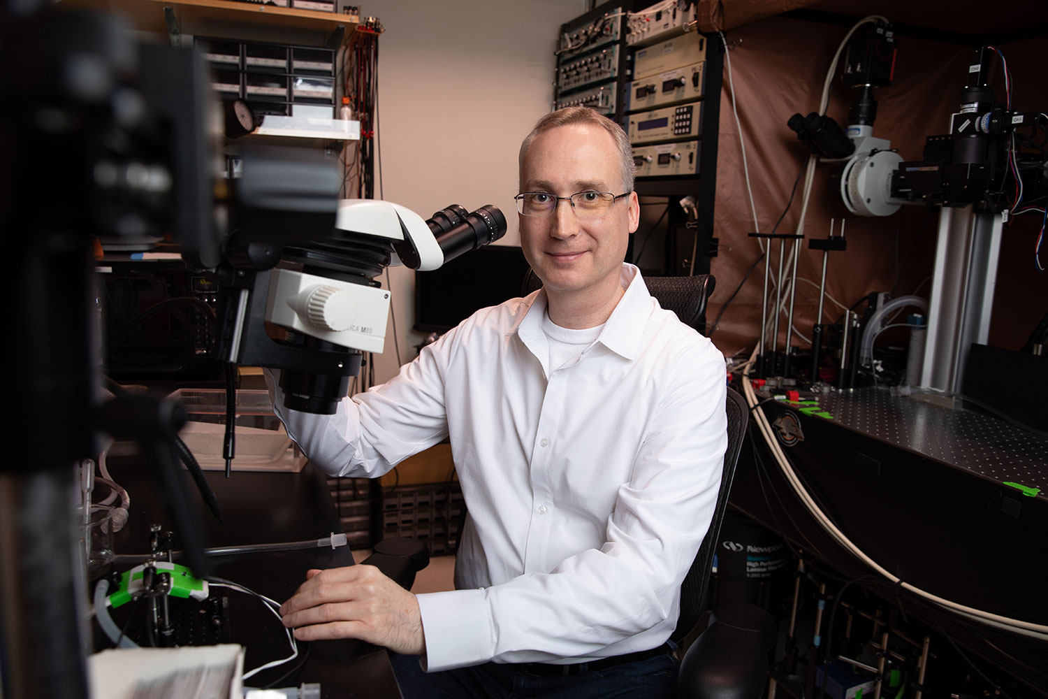
“This work establishes a versatile platform for imaging proteins with high specificity and minimal background,” says co-corresponding author Axel Nimmerjahn, PhD, professor and Françoise Gilot-Salk Chair at Salk. “It opens new opportunities to study how molecular and cellular processes unfold in real time across diverse biological systems.”
The innovation began with tiny protein fragments called nanobodies, which can be engineered to bind specific protein targets in living cells. When fused to fluorescent proteins, these nanobody-based probes can reveal where target proteins are located and how they behave. However, conventional versions can still generate signal even when unbound, creating background fluorescence that can obscure fine details.
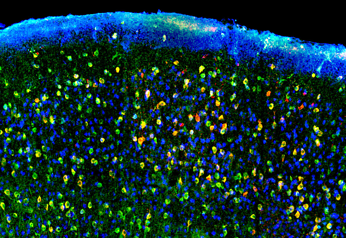
The Salk and Einstein team designed a new type of probe that retains the targeting power of nanobodies while greatly reducing background fluorescence. VIS-Fbs become stable and fluorescent only when bound to their intended target. This binding-dependent (“on-demand”) fluorescence reduces background noise by up to about a hundredfold, enabling much sharper visualization of protein location and dynamics.
In addition, the researchers developed multiple versions of this new probe that fluoresce across nearly the entire visible spectrum, from blue to far red. With this many color options, multiple cellular targets can be tracked simultaneously. Certain VIS-Fb variants can also be switched “on” and “off” with light, making it possible to follow protein behavior over time with high spatial and temporal precision. The researchers also established a modular design framework, enabling rapid adaptation of VIS-Fb probes to different targets and functional readouts.
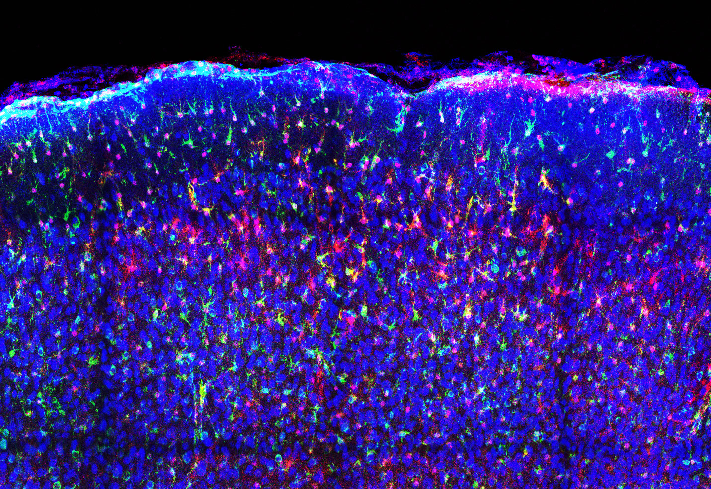
The new technology will allow scientists to gain more accurate, timely insight into cellular activity—even in complex environments like living brain tissue. The researchers demonstrated VIS-Fbs’ capabilities across a range of living models.
In mouse models, VIS-Fb probes enabled selective labeling and ratiometric imaging of calcium activity in neurons and astrocytes during behavior. In zebrafish, the technology allowed real-time tracking of dynamic changes during early development and in response to drugs that alter signaling pathways.
“Our results show that this imaging platform offers a much clearer and more precise view of how proteins behave inside living systems,” says co-corresponding author of the study Vladislav Verkhusha, PhD, professor and co-director of the Gruss Lipper Biophotonics Center at Albert Einstein College of Medicine. “It opens the door to studying complex biological processes, such as cell signaling, development, and disease progression, in new ways.”
Other authors include Erin Carey of Salk; Natalia Barykina, Juliana Mendonça-Gomes, and Sofia de Oliveira of the Albert Einstein College of Medicine; and Olena Oliinyk of the University of Helsinki.
This study was funded by the National Institutes of Health (GM122567, NS123719, GM147416), Jane and Aatos Erkko Foundation, Research Council of Finland, Finland Cancer Foundation, Chan Zuckerberg Initiative Foundation, NOMIS Foundation (Salk’s Neuroimmunology Initiative), and Edwards-Yeckel Research Foundation.
DOI: 10.1038/s41592-026-03056-3
JOURNAL
Natur Methoden
AUTOREN
Natalia V. Barykina, Erin M. Carey, Olena S. Oliinyk, Juliana M. Mendonça-Gomes, Sofia de Oliveira, Axel Nimmerjahn, and Vladislav V. Verkhusha
Büro für Kommunikation
Telefon: (858) 453-4100
press@salk.edu
Das Salk Institute ist ein unabhängiges, gemeinnütziges Forschungsinstitut, das 1960 von Jonas Salk, dem Entwickler des ersten sicheren und wirksamen Polio-Impfstoffs, gegründet wurde. Die Aufgabe des Instituts besteht darin, grundlegende, kooperative und risikofreudige Forschung voranzutreiben, die sich mit den dringendsten Herausforderungen der Gesellschaft befasst, darunter Krebs, Alzheimer und die Gefährdung der Landwirtschaft. Diese Grundlagenforschung bildet die Basis für alle translationalen Bemühungen und führt zu Erkenntnissen, die neue Medikamente und Innovationen weltweit ermöglichen.


