
June 15, 2016
Salk scientists show how T-cell receptors reposition during an immune response, revealing more on how the immune system is regulated
Salk scientists show how T-cell receptors reposition during an immune response, revealing more on how the immune system is regulated
LA JOLLA—When the body is fighting an invading pathogen, white blood cells—including T cells—must respond. Now, Salk Institute researchers have imaged how vital receptors on the surface of T cells bundle together when activated.
This study, the first to visualize this process in lymph nodes, could help scientists better understand how to turn up or down the immune system’s activity to treat autoimmune diseases, infections or even cancer. The results were published the week of June 13, 2016 in 美国国家科学院院刊.
Click-here 用于高分辨率图像。.
版权:萨克研究所
“We had seen these receptors cluster and reposition in cultured cells that were artificially stimulated in the lab, but we’ve never seen their natural arrangements in lymph nodes until now,” says senior author Björn Lillemeier, an associate professor in Salk’s Nomis Laboratories for Immunobiology and Microbial Pathogenesis, and the 韦特先进生物光子学中心.
T cells are activated when receptors embedded in their outer membrane bind to other immune cells that have digested an antigen, such as a virus, bacteria or 癌症 cell. In turn, the activated T cells switch on cellular pathways that help the body both actively seek out and destroy the antigen and remember it for the future. In the past, by looking at T-cell receptors embedded in isolated cells under the microscope, researchers discovered that the receptors are arranged in clusters—called protein islands—that merge when the cells are activated.
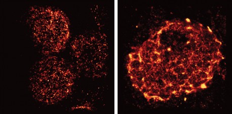
Lillemeier wanted more detail on how the receptors are arranged in tissue and how that arrangement might change when the T cells are activated in living hosts. The team used a super-resolution microscope developed in the laboratory of co-senior author Hu Cang, assistant professor at Salk’s Waitt Advanced Biophotonics Center and holder of the Frederick B. Rentschler Developmental Chair. This microscopy approach, called light-sheet direct stochastic optical reconstruction microscopy (dSTORM), let the researchers watch T cell receptors in the membranes of T cells in mouse lymph nodes at a resolution of approximately 50 nanometers.
The new imagery confirmed the previous observation that protein islands of T-cell receptors merge into larger “microclusters” when T cells are activated. But it also showed that, before cells are activated, the protein islands are already arranged in groups—dubbed “territories” by Lillemeier’s team. “The pre-organization on the molecular level basically turns the T cell into a loaded gun,” says Lillemeier.
Click here for a high-resolution image
版权:萨克研究所
The organization of surface receptors enables T cells to launch fast and effective immune response against antigens. Understanding how the molecular organization mediates the sensitivity of T cell responses could help researchers make the immune system more or less sensitive. In the case of autoimmune diseases, clinicians would like to turn down the immune system’s activity, while turning up the activity could help fight infections or cancers.
The research could also have implications for understanding other receptors in the body, which have a wide range of functions both within and outside the immune system. “We think that most receptors on the surfaces of cells are organized like this,” says Ying Hu, first author and postdoctoral researcher at the Salk Institute.
这项工作以及参与研究的学者得到了以下机构资助: NOMIS Foundation, , 那个 Waitt Foundation, the James B. Pendleton Charitable Trust, the 美国国立卫生研究院, , 那个 National Institute of Neurologic Disorders and Stroke, , 那个 国家癌症研究所 和 加州再生医学研究所.
日记
美国国家科学院院刊
作者
Ying S. Hu, Hu Cang, and Björn F. Lillemeier of the Salk Institute
宣传办公室
电话:(858) 453-4100
press@salk.edu
萨尔克研究所是一个独立的非营利性研究机构,由首个安全有效的脊髓灰质炎疫苗的研发者乔纳斯·索尔克于1960年创立。该研究所的使命是推动以合作、敢于冒险为特点的基础性研究,以应对癌症、阿尔茨海默病和农业脆弱性等社会最紧迫的挑战。这项基础科学支撑着所有的转化研究,产生有助于全球新药和创新的见解。.


