
September 15, 2015
New technique to selectively and noninvasively turn on groups of neurons in worms could be boon to science and medicine
New technique to selectively and noninvasively turn on groups of neurons in worms could be boon to science and medicine
LA JOLLA–Salk scientists have developed a new way to selectively activate brain, heart, muscle and other cells using ultrasonic waves. The new technique, dubbed sonogenetics, has some similarities to the burgeoning use of light to activate cells in order to better understand the brain.
This new method–which uses the same type of waves used in medical sonograms–may have advantages over the light-based approach–known as optogenetics–particularly when it comes to adapting the technology to human therapeutics. It was described September 15, 2015 in the journal Nature Communications.
“Light-based techniques are great for some uses and I think we’re going to continue to see developments on that front,” says 斯里坎特·查拉萨尼, an assistant professor in Salk’s 分子神经生物学实验室 and senior author of the study. “But this is a new, additional tool to manipulate neurons and other cells in the body.”
In optogenetics, researchers add light-sensitive channel proteins to neurons they wish to study. By shining a focused laser on the cells, they can selectively open these channels, either activating or silencing the target neurons. But using an optogenetics approach on cells deep in the brain is difficult: typically, researchers have to perform surgery to implant a fiber optic cable that can reach the cells. Plus, light is scattered by the brain and by other tissues in the body.
Chalasani and his group decided to see if they could develop an approach that instead relied on ultrasound waves for the activation. “In contrast to light, low-frequency ultrasound can travel through the body without any scattering,” he says. “This could be a big advantage when you want to stimulate a region deep in the brain without affecting other regions,” adds Stuart Ibsen, a postdoctoral fellow in the Chalasani lab and first author of the new work.
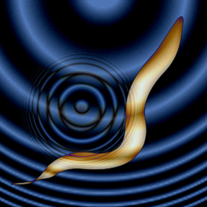
For the first time, sound waves are used to control brain cells. Salk scientists developed the new technique, dubbed sonogenetics, to selectively and noninvasively turn on groups of neurons in worms that could be a boon to science and medicine.
点击此处 用于高分辨率图像。.
图片:由萨克生物研究所提供
Chalasani and his colleagues first showed that, in the nematode Caenorhabditis elegans, microbubbles of gas outside of the worm were necessary to amplify the low-intensity ultrasound waves. “The microbubbles grow and shrink in tune with the ultrasound pressure waves,” Ibsen says. “These oscillations can then propagate noninvasively into the worm.”
Next, they found a membrane ion channel, TRP-4, which can respond to these waves. When mechanical deformations from the ultrasound hitting gas bubbles propagate into the worm, they cause TRP-4 channels to open up and activate the cell. Armed with that knowledge, the team tried adding the TRP-4 channel to neurons that don’t normally have it.
With this approach, they successfully activated neurons that don’t usually react to ultrasound.
So far, sonogenetics has only been applied to 秀丽隐杆线虫 neurons. But TRP-4 could be added to any calcium-sensitive cell type in any organism including humans, Chalasani says. Then, microbubbles could be injected into the bloodstream, and distributed throughout the body–an approach already used in some human imaging techniques. Ultrasound could then noninvasively reach any tissue of interest, including the brain, be amplified by the microbubbles, and activate the cells of interest through TRP-4. And many cells in the human body, he points out, can respond to the influxes of calcium caused by TRP-4.
“The real prize will be to see whether this could work in a mammalian brain,” Chalasani says. His group has already begun testing the approach in mice. “When we make the leap into therapies for humans, I think we have a better shot with noninvasive sonogenetics approaches than with optogenetics.”
Both optogenetics and sonogenetics approaches, he adds, hold promise in basic research by letting scientists study the effect of cell activation. And they also may be useful in therapeutics through the activation of cells affected by disease. However, for either technique to be used in humans, researchers first need to develop safe ways to deliver the light or ultrasound-sensitive channels to target cells.
Other researchers on the study were Stuart Ibsen and Ada Tong of the Salk Institute, and Carolyn Schutt and Sadik Esener of the 加州大学圣地亚哥分校.
The work and the researchers involved were supported by a Salk Institute Pioneer Fund Postdoctoral Fellowship, a Salk Institute Innovation Grant, the 丽塔·艾伦基金会, , 那个 W.M. Keck Foundation 和 美国国立卫生研究院.
日记
Nature Communications
作者
Stuart Ibsen, Ada Tong, Carolyn Schutt, Sadik Esener and Sreekanth H. Chalasani
宣传办公室
电话:(858) 453-4100
press@salk.edu
萨尔克研究所是一个独立的非营利性研究机构,由首个安全有效的脊髓灰质炎疫苗的研发者乔纳斯·索尔克于1960年创立。该研究所的使命是推动以合作、敢于冒险为特点的基础性研究,以应对癌症、阿尔茨海默病和农业脆弱性等社会最紧迫的挑战。这项基础科学支撑着所有的转化研究,产生有助于全球新药和创新的见解。.


