
June 15, 2016
Salk scientists show how T-cell receptors reposition during an immune response, revealing more on how the immune system is regulated
Salk scientists show how T-cell receptors reposition during an immune response, revealing more on how the immune system is regulated
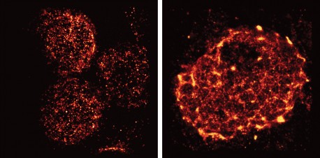
LA JOLLA—When the body is fighting an invading pathogen, white blood cells—including T cells—must respond. Now, Salk Institute researchers have imaged how vital receptors on the surface of T cells bundle together when activated.
This study, the first to visualize this process in lymph nodes, could help scientists better understand how to turn up or down the immune system’s activity to treat autoimmune diseases, infections or even cancer. The results were published the week of June 13, 2016 in Verhandlungen der Nationalen Akademie der Wissenschaften.
Click-here für ein hochauflösendes Bild.
Kredit: Salk Institut
“We had seen these receptors cluster and reposition in cultured cells that were artificially stimulated in the lab, but we’ve never seen their natural arrangements in lymph nodes until now,” says senior author Björn Lillemeier, an associate professor in Salk’s Nomis Laboratories for Immunobiology and Microbial Pathogenesis, and the Waitt Advanced Biophotonics Center.
T cells are activated when receptors embedded in their outer membrane bind to other immune cells that have digested an antigen, such as a virus, bacteria or Krebs cell. In turn, the activated T cells switch on cellular pathways that help the body both actively seek out and destroy the antigen and remember it for the future. In the past, by looking at T-cell receptors embedded in isolated cells under the microscope, researchers discovered that the receptors are arranged in clusters—called protein islands—that merge when the cells are activated.
Lillemeier wanted more detail on how the receptors are arranged in tissue and how that arrangement might change when the T cells are activated in living hosts. The team used a super-resolution microscope developed in the laboratory of co-senior author Hu Cang, assistant professor at Salk’s Waitt Advanced Biophotonics Center and holder of the Frederick B. Rentschler Developmental Chair. This microscopy approach, called light-sheet direct stochastic optical reconstruction microscopy (dSTORM), let the researchers watch T cell receptors in the membranes of T cells in mouse lymph nodes at a resolution of approximately 50 nanometers.
The new imagery confirmed the previous observation that protein islands of T-cell receptors merge into larger “microclusters” when T cells are activated. But it also showed that, before cells are activated, the protein islands are already arranged in groups—dubbed “territories” by Lillemeier’s team. “The pre-organization on the molecular level basically turns the T cell into a loaded gun,” says Lillemeier.
Click here for a high-resolution image
Kredit: Salk Institut
The organization of surface receptors enables T cells to launch fast and effective immune response against antigens. Understanding how the molecular organization mediates the sensitivity of T cell responses could help researchers make the immune system more or less sensitive. In the case of autoimmune diseases, clinicians would like to turn down the immune system’s activity, while turning up the activity could help fight infections or cancers.
The research could also have implications for understanding other receptors in the body, which have a wide range of functions both within and outside the immune system. “We think that most receptors on the surfaces of cells are organized like this,” says Ying Hu, first author and postdoctoral researcher at the Salk Institute.
The work and the researchers involved were supported by grants from the NOMIS Foundation, der Waitt Foundation, the James B. Pendleton Charitable Trust, the Nationale Gesundheitsinstitute, der National Institute of Neurologic Disorders and Stroke, der National Cancer Institute und die California Institute for Regenerative Medicine.
JOURNAL
Verhandlungen der Nationalen Akademie der Wissenschaften
AUTOREN
Ying S. Hu, Hu Cang, and Björn F. Lillemeier of the Salk Institute
Büro für Kommunikation
Telefon: (858) 453-4100
press@salk.edu
Das Salk Institute ist ein unabhängiges, gemeinnütziges Forschungsinstitut, das 1960 von Jonas Salk, dem Entwickler des ersten sicheren und wirksamen Polio-Impfstoffs, gegründet wurde. Die Aufgabe des Instituts besteht darin, grundlegende, kooperative und risikofreudige Forschung voranzutreiben, die sich mit den dringendsten Herausforderungen der Gesellschaft befasst, darunter Krebs, Alzheimer und die Gefährdung der Landwirtschaft. Diese Grundlagenforschung bildet die Basis für alle translationalen Bemühungen und führt zu Erkenntnissen, die neue Medikamente und Innovationen weltweit ermöglichen.


